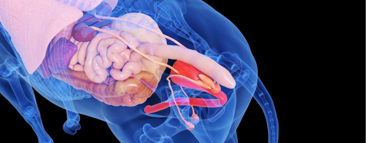
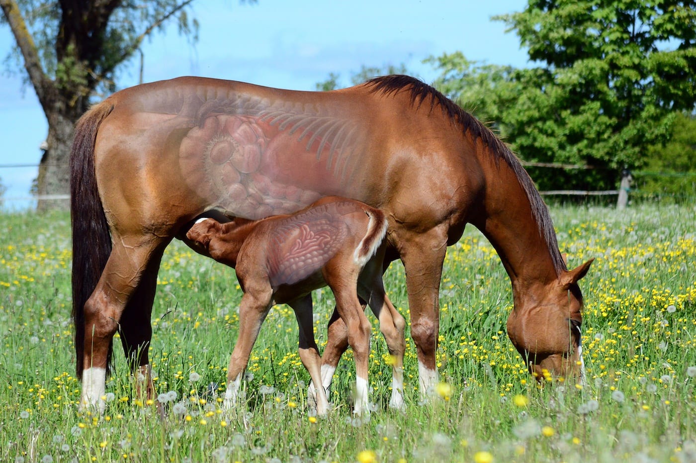
Horses have a large and complex digestive system susceptible to multiple health concerns associated with modern management. Although many owners may assume that gastric ulcers are responsible for any gut-related discomfort their horse experiences, digestive health does not stop at the stomach.
Conditions in the horse’s hindgut are far more common than you may realize and can significantly impact overall health. Keep reading to learn how to identify these problems and implement management practices that help keep your horse comfortable by supporting a healthy hindgut.
The Importance of a Healthy Hindgut
Extensive research and modern treatments have emphasized equine gastric ulcers in recent years. But in reality, a horse’s stomach makes up less than 10% of the equine digestive tract’s total volume.
The hindgut includes the cecum and colon, the largest structures in the equine gastrointestinal tract. These structures also house a population of beneficial bacteria responsible for digesting fiber, the primary energy source in the equine diet.
Fibrous material in the hindgut provides a water reservoir that promotes hydration, and bacterial fermentation of dietary fiber also produces essential vitamins absorbed by the horse. Although the full capabilities of equine hindgut bacteria are still unclear, the gut microbiota may also impact immune function and behavior.
Poor hindgi tract health and an unbalanced microbiota can prevent the absorption of vital dietary components necessary for peak health and performance. As a result, horses that struggle with common equine hindgut problems may experience dehydration, reduced appetite, poor hoof condition, impaired immune function, and attitude changes.
Problems in the Equine Hindgut
The equine digestive system evolved to thrive on continuous grazing. But today, the lifestyles of modern performance horses who live in stalls, face rigorous training and competition, and eat intermittently and often with high volumes of grain, often place them at risk for common hindgut problems.
Colonic Ulcers
Most horse people are familiar with gastric ulcers that occur in the foregut. But ulcers can also occur in the hindgut. Colonic ulcers involve a thinning in the colon lining and can be challenging to diagnose as the colon cannot be scoped the way the stomach can be.
Ulcers have different severities depending on the thickness of the colon’s outer wall. However, surgery is the only way to examine an ulcer in the hindgut thoroughly. Since surgery is typically avoided, veterinarians mostly rely on ultrasound, bloodwork, and clinical signs.
Although stomach ulcers are more common, examinations during necropsies revealed that 44 to 60 percent of horses have some colonic ulceration (Pellegrini, 2005). Clinical signs such as chronic colic, lethargy, or diarrhea are typically associated with this hindgut issue.
Hindgut Acidosis
Colonic ulcers are also frequently associated with hindgut acidosis. This condition occurs when starch overloads the digestive system, causing drastic changes to the pH of the hindgut. This excessive acidity often results from a high grain and low forage diet and impacts nearly 60% of all performance horses.
Enzymes in the small intestine digest dietary starch. But large meals with high volumes of grain can exceed the horse’s capacity to absorb all of the starch. When this happens, undigested starch can move into the large intestine, where bacterial fermentation produces increased lactic acid. This increase in lactic acid causes a sharp decline in pH, which can lead to a change in the bacterial population there.
Fiber-fermenting bacteria are less efficient in acidic environments. These bacteria begin dying off and releasing endotoxins that can enter the bloodstream through a weakened mucosal lining and initiate an inflammatory response. This inflamation increases susceptibility to laminitis and colic (Milinovich et al., 2007).
Digestive Imbalance
Digestive imbalance or dysbiosis involves a significant imbalance in the intestinal microbiota of the digestive tract. This condition can result in colitis, laminitis, or colic. But even small changes in this delicate ecosystem can have big health implications.
A healthy microbiota is essential for digesting the fibrous diet horses evolved to eat. Bacteria ferment fiber in the hindgut to create volatile fatty acids that horses use for energy. The microbial population also produces essential vitamins for the horse to absorb (Julliand and Grim, 2016).
Digestive imbalances can occur due to sudden dietary changes, grain overload, or antibiotic use. Clinical signs of dysbiosis depend on the area of the intestine affected but often include colic or systemic inflammation.
Colitis
Colitis describes any inflammation of the lining of the large intestine. This inflammation is actually a set of symptoms associated with multiple infectious and non-infectious causes. To treat colitis effectively, the veterinarian must identify the underlying cause.
Infectious agents of colitis include some parasites, certain viruses, and bacterial diseases like Potomac Horse Fever. Non-infectious causes include diarrhea from antibiotics, sand irritation, dietary imbalances, inflammatory bowel disease, toxicities, or NSAID toxicosis (Larsen, 1997).
Colitis can have severe implications, including dehydration from diarrhea and compromised nutrition from reduced nutrient absorption.
Right Dorsal Colitis
Right Dorsal Colitis is a specific type of ulceration found in the upper right portion of the equine colon. This condition has a solid link to the use of non-steroidal anti-inflammatory drugs or NSAIDs, such as phenylbutazone (Collins and Tyler, 1985).
NSAIDs are commonly used to address aches and pains in horses. Still, long-term use of these drugs at high doses can reduce blood flow to the mucosa, leaving the sensitive upper right section of the colon susceptible to ulceration.
Intestinal mucosa damage can also cause water leakage into the bowel and low blood protein. However, NSAID toxicosis is not just associated with hindgut problems alone. This condition may also result in damage to the stomach, esophagus, mouth, and kidneys (Jones et al., 2009).
Diagnosing Problems in the Equine Hindgut
Colonic ulcers and other equine hindgut conditions can be incredibly challenging to identify. Common symptoms associated with hindgut problems often resemble classic clinical signs of other digestive issues. In addition, behavior problems that seem to be training issues initially may also indicate hindgut discomfort.
Early clinical signs that your horse may have a hindgut problem include recurring colic, loss of appetite, lethargy, diarrhea, and sensitivity along his flanks. Before you begin a treatment plan for gastric ulcers, ask your veterinarian to examine your horse for potential hindgut problems.
Gastroscopy can confirm ulcers in the stomach but can’t be used in the hindgut. So instead, most veterinarians will perform a transabdominal ultrasound if they suspect hindgut ulcers. The ultrasound allows the veterinarian to measure the colon’s outer layer and identify potential ulceration.
A fecal blood test like SUCCEED FBT is another tool that helps veterinarians identify hindgut lesions. This test detects the presence of albumin and hemoglobin in stool samples. These blood proteins can leak through a weakened intestinal lining and indicate the potential presence of hindgut damage or ulcers.
Support Hindgut Health For A Healthy Horse
The demands placed on modern performance horses can compromise hindgi tract health and contribute to troublesome digestive issues. Domestic horses also often have lifestyles and diets that differ significantly from their grazing ancestors, despite having similar digestive systems.
Modern equine diets with insufficient fiber and high volumes of simple carbohydrates can overwhelm the small intestine and cause imbalances in the hindgut. But simple management decisions like providing free-choice forage, increasing turnout time, feeding smaller meals, and limiting starch intake can help keep your horse’s hindgi tract healthy.
Daily digestive support is also often necessary for performance horses subject to the extra demands of travel, competition, and training. Look for products that support the health of the entire gastrointestinal tract by promoting a healthy microbiota and mucosal barrier with ingredients like oat oil, yeast, L-glutamine, and L-threonine.
Identify Hindgut Problems with SUCCEED FBT
Equine hindgut issues are more common than you may realize. Even if your horse has a confirmed diagnosis of gastric ulcers, your evaluation should not stop at the stomach.
The SUCCEED Equine Fecal Blood Test allows your veterinarian to identify hindgut lesions or other pathological conditions with an inexpensive and non-invasive stall-side screen test. The FBT uses antibodies to detect the presence of the blood protein albumin in a fecal sample, indicating a problem in the hindgut that your veterinarian can investigate further.
Proactively managing your horse’s hindgi tract health with appropriate feeding practices, daily digestive support, and veterinary diagnostic tools can ensure that your horse stays comfortable enough to perform at his best. If you suspect your horse may be struggling with hindgut problems, ask your vet to test your horse with the SUCCEED FBT today.
References
- Pellegrini, F.L. (2005) Results of a large-scale necroscopic study of equine colonic ulcers. Journal of Equine Veterinary Science 25:113-117.
- Milinovich, G.J., et al. (2007) Fluorescence in situ hybridization analysis of hindgut bacteria associated with the development of equine laminitis. Environ. Microbiol. 9(8): 2090–2100.
- Julliand, V. and Grimm, P. (2016) Horse Species Symposium: The microbiome of the horse hindgut: History and current knowledge. Journal of Animal Science.
- Larsen, J. (1997) Acute colitis in adult horses. A review with emphasis on aetiology and pathogenesis. Vet Q 19 (2): 72–80
- Collins, L.G., Tyler, D.E. (1985) Experimentally induced phenylbutazone toxicosis in ponies: description of the syndrome and its prevention with synthetic prostaglandin E2. Am J Vet Res. 46(8): 1605-15
- Jones, S.L. et al. (2009) Right Dorsal Colitis. In: Current Therapy in Equine Medicine, ed. N.E. Robinson and K.A. Sprayberry. Elsevier pp. 430-432.


